Biobite_Etco2

What is EtCO₂?

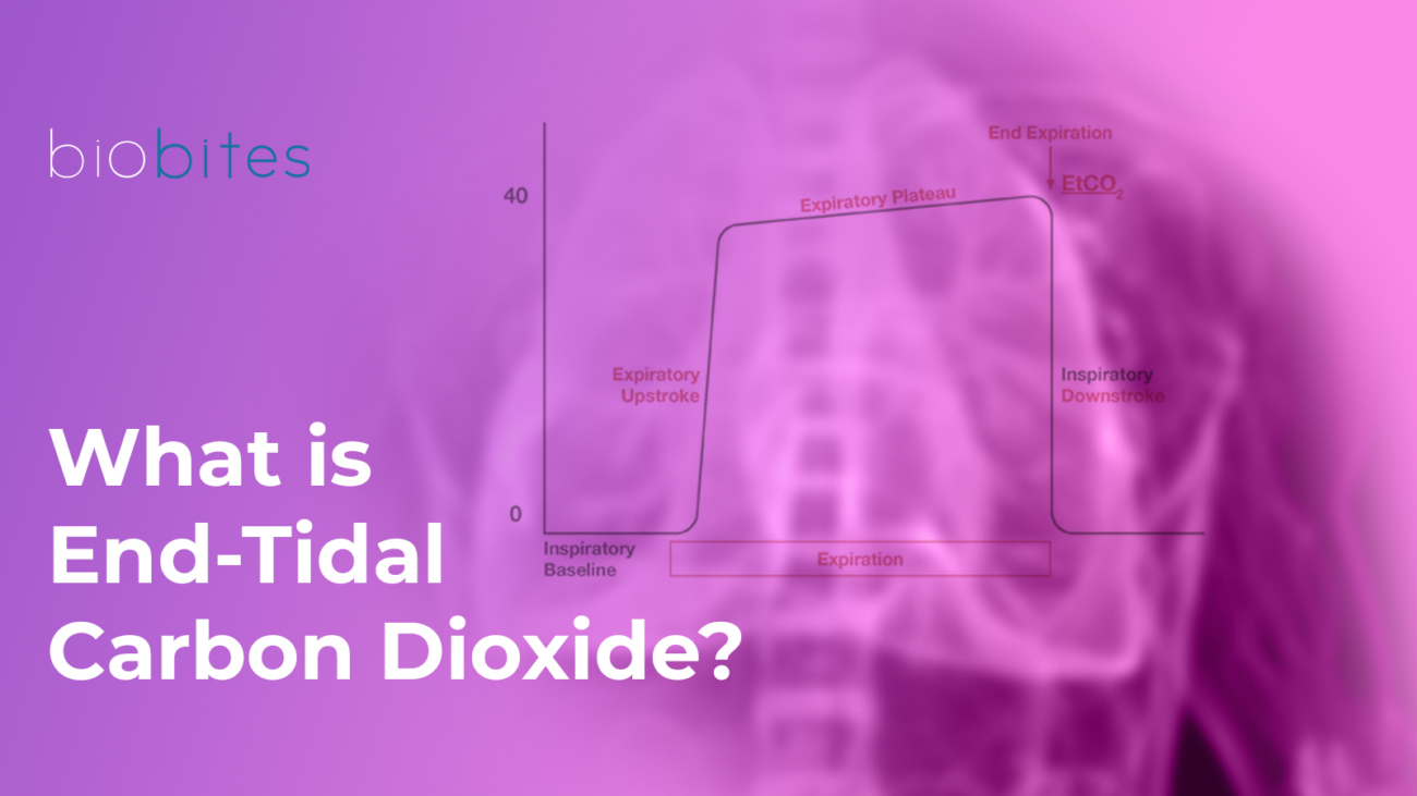
EtCO₂ (End-Tidal Carbon Dioxide) refers to the measurement of the carbon dioxide concentration in exhaled air at the end of expiration. It is typically expressed in millimeters of mercury (mmHg) and measured using a capnography device. It provides critical insights into the respiratory system, cardiac circulation, and metabolism.

At the end of the respiratory cycle, during exhalation, EtCO₂ represents the peak concentration of carbon dioxide (CO₂) in the exhaled gas. This value forms the basis of EtCO₂ monitoring. By reflecting the interaction between the respiratory system, circulation, and metabolism, it plays a crucial role in assessing the patient’s overall condition.

Normal EtCO₂ Values

The normal EtCO₂ level typically ranges between 35 and 45 mmHg.

  • Low EtCO₂: May indicate conditions such as hyperventilation, low cardiac output, or pulmonary embolism.
  • High EtCO₂: May suggest hypoventilation, excessive metabolic activity, or inadequate ventilator settings.

Monitoring EtCO₂ levels with mechanical ventilators plays a critical role in evaluating a patient’s respiratory status and the effectiveness of ventilation. This monitoring is performed using capnography devices, which are often integrated into the ventilator system.

EtCO₂ Monitoring Methods

  1. Mainstream Capnography
    • How It Works: The sensor is placed between the endotracheal or tracheostomy tube and the breathing circuit. The patient’s exhaled air passes directly through the sensor, and CO₂ concentration is measured in real-time.
    • Advantages: Provides instantaneous and accurate measurements with minimal delay during exhalation.
    • Disadvantages: The sensor’s weight and heat may cause discomfort, particularly for small children or frail patients.
  2. Sidestream Capnography
    • How It Works: A small gas sample is drawn from the breathing circuit and delivered to the analyzer within the device. This method can also be used for non-intubated patients.
    • Advantages: Compatible with various breathing circuits and suitable for patients who do not require intubation.
    • Disadvantages: Condensation may accumulate in the sampling line, potentially affecting measurement accuracy.

Ventilation Efficiency Through EtCO₂ Monitoring

EtCO₂ monitoring is a direct method to evaluate ventilation efficiency. It demonstrates how effectively the respiratory system eliminates carbon dioxide (CO₂) and assesses the efficiency of CO₂ removal produced through metabolic processes via ventilation. Ventilation efficiency is evaluated in various clinical conditions using EtCO₂ levels and capnogram analyses.

  1. Hypoventilation (Decreased Ventilation):
    • EtCO₂ Level: Increased (typically > 45 mmHg).
    • Cause: Inadequate elimination of CO₂ by the lungs.
    • Clinical Conditions: Respiratory depression, sedation, neuromuscular blockade, obesity hypoventilation syndrome.
    • Effect: Elevated EtCO₂ levels indicate the need to enhance ventilation.
  2. Hyperventilation (Increased Ventilation):
    • EtCO₂ Level: Decreased (typically < 35 mmHg).
    • Cause: Excessive elimination of CO₂.
    • Clinical Conditions: Anxiety, pain, response to hypoxia, compensatory mechanisms.
    • Effect: A reduction in ventilation rate or tidal volume may be necessary.
  3. Increased Alveolar Dead Space:
    • EtCO₂ Level: Decreased, but arterial CO₂ (PaCO₂) remains elevated.
    • Cause: Reduced pulmonary perfusion or ventilation-perfusion (V/Q) mismatch.
    • Clinical Conditions: Pulmonary embolism, low cardiac output, shock.
    • Effect: The difference between EtCO₂ and PaCO₂ increases (EtCO₂-PaCO₂ gradient).
  4. Circulatory Failure:
    • EtCO₂ Level: Decreased.
    • Cause: Reduced cardiac output decreases the amount of CO₂ delivered to the alveoli.
    • Clinical Conditions: Cardiac arrest, low-perfusion shock.
    • Effect: Used to monitor the effectiveness of CPR; an increase in EtCO₂ indicates the restoration of circulation.

FAQs

  1. What is the normal EtCO₂ value?
    The normal range is between 35-45 mmHg.
  2. What does low EtCO₂ mean?
    Low EtCO₂ refers to a condition where the carbon dioxide level in exhaled air is below normal. This may indicate issues related to ventilation, perfusion, or metabolism. EtCO₂ levels below 35 mmHg are typically referred to as hypocapnia.
  3. What are the causes of low EtCO₂ on a ventilator?
    Low EtCO₂ is commonly caused by factors such as hyperventilation, hypoperfusion, airway issues, or decreased metabolic activity.

References

  • Aminiahidashti, Hamed, et al. “Applications of end-tidal carbon dioxide (ETCO2) monitoring in emergency department; a narrative review.” Emergency 6.1 (2018).
  • Trilĺo, Giulio, Martin von Planta, and Fulvio Kette. “ETCO2 monitoring during low flow states: clinical aims and limits.” Resuscitation 27.1 (1994): 1-8.
  • Miner, James R., William Heegaard, and David Plummer. “End‐tidal carbon dioxide monitoring during procedural sedation.” Academic Emergency Medicine 9.4 (2002): 275-280.
  • Paiva, Edison F., James H. Paxton, and Brian J. O’Neil. “The use of end-tidal carbon dioxide (ETCO2) measurement to guide management of cardiac arrest: a systematic review.” Resuscitation 123 (2018): 1-7.
Biobite_First_1 (1)

What is Intraoperative Neuromonitoring?

Intraoperative neuromonitoring (IONM) is a technique aimed at monitoring and protecting the nervous system, particularly nerves and the spinal cord, during surgery. It is primarily used in surgeries where there is a high risk of damage to the nerves or nervous system.

Why is intraoperative neuromonitoring important?

Neuromonitor reduces the risk of permanent damage by enabling real-time monitoring of nerve functions during surgeries that may pose a threat to the nervous system. Neuromonitoring devices facilitate the surgeon’s work by identifying potential damage to nerves in procedures involving the brain, spinal cord, and nerve roots, thus providing the capability to monitor and protect nerve tissues. For instance, the use of IONM in brain tumor and spinal surgeries helps prevent complications such as sensory loss or paralysis.

Additionally, nerve monitoring can positively impact the postoperative recovery process. By minimizing permanent losses in motor and sensory functions, IONM contributes to an improved quality of life for the patient.

The Benefits of Neuromonitoring on Patient Outcomes

 Neuromonitoring offers significant health and safety benefits by enabling real-time monitoring of patients’ nervous systems during surgical procedures. Neuromonitoring devices are commonly used in brain, spinal, vascular, and thyroid surgeries, helping to minimize surgical risks by preventing damage to nerve tissues.

Surgeries Where Neuromonitoring Devices Are Commonly Used

  • Thyroid & Parathyroid Surgeries: Neuromonitoring is a crucial technology in preventing nerve injuries, particularly focusing on protecting the nerves that control the vocal cords. These nerves are located very close to the thyroid gland, and any damage to them during surgery can lead to complications such as hoarseness or even loss of voice. IONM allows the surgeon to work with precision by monitoring these nerves, thereby reducing the risk of nerve injury. The use of IONM is especially beneficial in recurrent goiter and complex thyroid surgeries, as it helps minimize issues like hoarseness and accelerates the postoperative recovery process.
  • Spinal Surgeries: In spinal surgery, IONM plays an essential role in protecting nerve roots, the spinal cord, and neural pathways. Spinal surgeries carry a high risk of nerve or spinal cord injury, necessitating a monitoring system to help the surgeon perform the procedure safely.
  • Brain Surgeries: Brain surgeries are performed in areas containing delicate neural tissues, and the use of neuromonitoring devices helps protect neurological structures by monitoring the brain and nervous system in real time.
  • Peripheral Nerve Surgeries: Peripheral nerves transmit sensory and motor signals to specific parts of the body. Damage to these nerves can lead to complications such as loss of muscle control, sensory disturbances, or pain. In peripheral nerve surgeries, neuromonitoring devices provide the surgeon with real-time information to prevent nerve damage, ensuring effective nerve monitoring.
  • Vascular Surgeries: Neuromonitoring is employed when nerves are located close to vascular structures and there is a risk of nerve injury. In surgeries on major blood vessels, protecting nerves around these structures is critical. For example, procedures like carotid artery surgery or aortic aneurysm repairs carry a high risk of nerve damage, making neuromonitoring essential for safeguarding these nerves.

FAQs

1. What are the risks of not using neuromonitoring in certain surgeries?

In surgeries where neuromonitoring is not used, the risk of nerve damage increases, which can lead to temporary or permanent neurological complications. When nerves are unprotected, patients may experience permanent paralysis, sensory loss, or other motor and sensory impairments.

2. Who performs and monitors IONM during surgery?

The neuromonitoring setup is managed by a neuromonitoring specialist, and it is used collaboratively by the surgeon and anesthesiologist throughout the surgery.

3. Is intraoperative neuromonitoring safe for all patients?

While IONM is generally considered a safe technique, the electrodes in contact with the skin can cause allergic reactions or skin irritation in some patients. Temporary side effects such as localized pain or muscle spasms may also occur in certain cases.

References:

  1. Skinner, S.A. ∙ Cohen, B.A. ∙ Morledge, D.E. Practice guidelines for the supervising professional: intraoperative neurophysiological monitoring. J Clin Monit Comput. 2014; 28:103-111
  2. R Malik, D Linos. Intraoperative neuromonitoring in thyroid surgery: a systematic review. World journal of surgery, 2016 – Springer
  3.  Wong, Andrew K., et al. “Intraoperative neuromonitoring.” Neurologic Clinics 40.2 (2022): 375-389.
  4. Shils, Jay L., and Tod B. Sloan. “Intraoperative neuromonitoring.” International anesthesiology clinics 53.1 (2015): 53-73.