Biobite_First_1 (1)

What is Intraoperative Neuromonitoring?

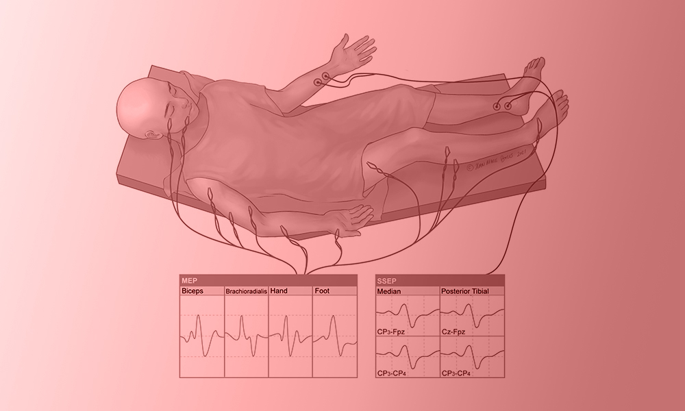
Intraoperative neuromonitoring (IONM) is a technique aimed at monitoring and protecting the nervous system, particularly nerves and the spinal cord, during surgery. It is primarily used in surgeries where there is a high risk of damage to the nerves or nervous system.

Why is intraoperative neuromonitoring important?

Neuromonitor reduces the risk of permanent damage by enabling real-time monitoring of nerve functions during surgeries that may pose a threat to the nervous system. Neuromonitoring devices facilitate the surgeon’s work by identifying potential damage to nerves in procedures involving the brain, spinal cord, and nerve roots, thus providing the capability to monitor and protect nerve tissues. For instance, the use of IONM in brain tumor and spinal surgeries helps prevent complications such as sensory loss or paralysis.

Additionally, nerve monitoring can positively impact the postoperative recovery process. By minimizing permanent losses in motor and sensory functions, IONM contributes to an improved quality of life for the patient.

The Benefits of Neuromonitoring on Patient Outcomes

 Neuromonitoring offers significant health and safety benefits by enabling real-time monitoring of patients’ nervous systems during surgical procedures. Neuromonitoring devices are commonly used in brain, spinal, vascular, and thyroid surgeries, helping to minimize surgical risks by preventing damage to nerve tissues.

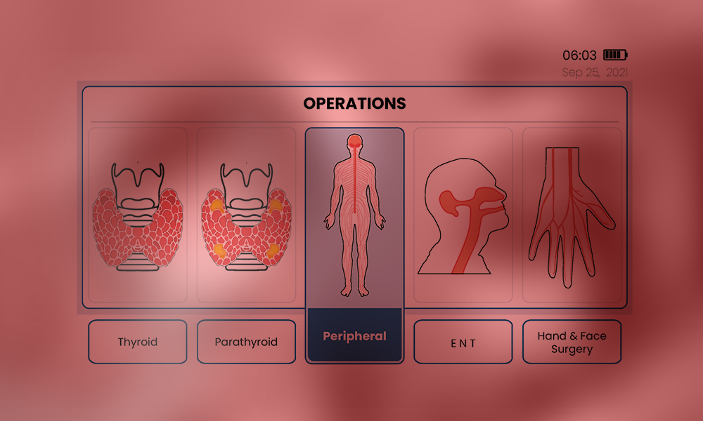
Surgeries Where Neuromonitoring Devices Are Commonly Used

  • Thyroid & Parathyroid Surgeries: Neuromonitoring is a crucial technology in preventing nerve injuries, particularly focusing on protecting the nerves that control the vocal cords. These nerves are located very close to the thyroid gland, and any damage to them during surgery can lead to complications such as hoarseness or even loss of voice. IONM allows the surgeon to work with precision by monitoring these nerves, thereby reducing the risk of nerve injury. The use of IONM is especially beneficial in recurrent goiter and complex thyroid surgeries, as it helps minimize issues like hoarseness and accelerates the postoperative recovery process.
  • Spinal Surgeries: In spinal surgery, IONM plays an essential role in protecting nerve roots, the spinal cord, and neural pathways. Spinal surgeries carry a high risk of nerve or spinal cord injury, necessitating a monitoring system to help the surgeon perform the procedure safely.
  • Brain Surgeries: Brain surgeries are performed in areas containing delicate neural tissues, and the use of neuromonitoring devices helps protect neurological structures by monitoring the brain and nervous system in real time.
  • Peripheral Nerve Surgeries: Peripheral nerves transmit sensory and motor signals to specific parts of the body. Damage to these nerves can lead to complications such as loss of muscle control, sensory disturbances, or pain. In peripheral nerve surgeries, neuromonitoring devices provide the surgeon with real-time information to prevent nerve damage, ensuring effective nerve monitoring.
  • Vascular Surgeries: Neuromonitoring is employed when nerves are located close to vascular structures and there is a risk of nerve injury. In surgeries on major blood vessels, protecting nerves around these structures is critical. For example, procedures like carotid artery surgery or aortic aneurysm repairs carry a high risk of nerve damage, making neuromonitoring essential for safeguarding these nerves.

FAQs

1. What are the risks of not using neuromonitoring in certain surgeries?

In surgeries where neuromonitoring is not used, the risk of nerve damage increases, which can lead to temporary or permanent neurological complications. When nerves are unprotected, patients may experience permanent paralysis, sensory loss, or other motor and sensory impairments.

2. Who performs and monitors IONM during surgery?

The neuromonitoring setup is managed by a neuromonitoring specialist, and it is used collaboratively by the surgeon and anesthesiologist throughout the surgery.

3. Is intraoperative neuromonitoring safe for all patients?

While IONM is generally considered a safe technique, the electrodes in contact with the skin can cause allergic reactions or skin irritation in some patients. Temporary side effects such as localized pain or muscle spasms may also occur in certain cases.

References:

  1. Skinner, S.A. ∙ Cohen, B.A. ∙ Morledge, D.E. Practice guidelines for the supervising professional: intraoperative neurophysiological monitoring. J Clin Monit Comput. 2014; 28:103-111
  2. R Malik, D Linos. Intraoperative neuromonitoring in thyroid surgery: a systematic review. World journal of surgery, 2016 – Springer
  3.  Wong, Andrew K., et al. “Intraoperative neuromonitoring.” Neurologic Clinics 40.2 (2022): 375-389.
  4. Shils, Jay L., and Tod B. Sloan. “Intraoperative neuromonitoring.” International anesthesiology clinics 53.1 (2015): 53-73.

Understanding Neuromonitoring

Understanding Neuromonitoring: Applications and Advancement

During an operation, surgeons often employ various activities such as intraoperative monitoring (IOM) to achieve the ideal goal of saving lives. However, in cases related to the nervous system an IOM subdivision process called Neuromonitoring or Electrophysiologic monitoring is often employed.

It provides a monitoring purpose that notifies the anesthesia specialist and operating neurologist of an approaching injury so that they can adjust the course of treatment in time to avoid irreversible harm.

This is sometimes used to create and locate neuron regions to help provide technical management. It is a strategy that is employed to reduce possible risks associated with the neural network.

Continue reading as this article provides you with information about the meaning of intraoperative neuromonitoring (IONM), its basics, clinical application, and its technical future directions.

Neuromonitor

What’s Intraoperative Neuromonitoring?

One of the medical procedures that entails constant checks of brain and neuronal activities during neural operations is known as intraoperative neuromonitoring (IONM). This procedure aids in the structural presentation of the sensorium and also illustrates its likely associated impairments.

Its main objective is to maintain the neurological system’s ideal nature, most especially the central nervous system (CNS) and the peripheral nerve. In addition, IONM provides neurophysiological data, which presents safer and more comprehensive processes that can be executed by the neurologist.

Basics of Neuromonitoring: What you need to know?

In many medical procedures, neuromonitoring has become the standard and has taken the place of intraoperative wake-up testing in spinal surgery. However, there are no evaluating terms and standards of care as most monitoring teams and surgeons choose their methods. Below are its basics:

  • Objective: During an operation, neuromonitoring is often used to obtain nerves’ electrical potentials produced by their axons. This is carried out to prevent and identify problems before escalating and to reduce any possible complications.
  • Methods: there are various methods used for surgical neuromonitoring. These are: 
  1. Electromyography (EMG): is used for recording muscular activities.
  2. Electroencephalography (EEG): is utilized for measuring brain activity.
  3. Motor Evoked Potential (MEP): This is often used for evaluating motor pathway function.
  4. Somatosensory Evoked Potentials (SSEP): To track sensory pathways integrity.
  • Application: Neuromonitoring in surgery is often applied in different ways to a wide range of neurological problems eg. Cranial neurosurgery, interventional radiological procedures, Orthopedic spinal correction, stroke, hypoxic-ischemic injury, meningitis, etc.
  • Procedure: To measure brain reactions,  perfectly placed electrodes are applied to the patient, and baseline values are recorded before the commencement of the surgical operation. Throughout the process, there is always proper evaluation, and a swift reaction is often taken whenever there is a baseline alteration.
  • Teamwork: There is always a cordial interaction between Neurologists, surgeons, and anesthetic teams to obtain current data about brain activity. With this obtained data, the surgical team needs to discuss possible steps and plans among themselves that will help in direct clinical decision-making.
  • Continuous Evaluation: As the operation continues, there is always a concurrent check that makes the surgeons quickly identify any possible variation in nerve function to make any necessary corrections.

Cutting-Edge Applications in Neuromonitoring Clinical Settings

With the rapid changes and developments in today’s neuromonitoring technology, significant advances have been made in the medical world. These modern techniques in clinical settings have improved immediate nervous system diagnosis and protection. Here are some conspicuous examples:

  • Advanced Intraoperative MRI: During surgeries, an intraoperative radiological imaging device provides structural or regional images that aid quick nervous detection by neurologists.  These magnetic resonated images facilitate easy access to various modifications in the brain or spinal cord.
  • Optical Coherence Tomography (OCT): The images of some complex optical regions like the optic nerve, and retina can be obtained at high quality. It has been put to use many times to diagnose and manage diabetes’s effect on the retina in conditions like glaucoma and retinopathy.
  • High-Resonance Image: The application of neuromonitoring in surgeries provides an exceptional method for surgeons to craft comprehensive preoperative planning. Most of the time, it grants fast access to complications during operations with its additional high-resonating imaging features.
  • Neural stimulation Technique: It comprises advanced methods that use magnetic or electrical stimulation to modify the neuronal response. Also, this technique is often used to map and protect important brain circuits.
  • Mechanical Alarming System: This is one of the most important innovative applications of surgical neuromonitoring that improves responsiveness. This system automatically identifies and notifies surgical teams of possible problems with the help of AI algorithms.
Neuromonitoring photo 2

Clinical Application of Neuromonitoring in Surgery

The application of surgical neuromonitoring methods involves a wide range of healthcare processes that improve patients’ outcomes. Some of its vital applications in medics are:

  • Surgeries on the brain: Neuromonitoring helps evaluate and protect vital cranial functions during brain surgeries, reducing the possibility of neurological implications. It is used during brain tumor removal and epileptic surgery.
  • Spinal Cord Surgery: Intraoperative neuromonitoring plays an exceptional role in countless surgeries related to the spine. Some of these are laminectomy, spinal decompression, and fusion. IONM also acts as a protective medium for the spine to evaluate nerve fibers during surgeries.
  • Musculoskeletal surgery: In conditions related to the skeletal system (joint, suture, or skull)  the use of neuromonitoring in surgery is of great significance. Some orthopedic surgeries like laminectomy and kyphoplasty are special operations where electrophysiology monitoring methods can be employed.
  • Complicated Pediatric Operations: The nervous system in children with complicated medical conditions such as scoliosis and spinal tumors, is monitored to avoid neuronal growth retardation.
  • Otolaryngological Operations: Intraoperative neuromonitoring helps maintain important neural structures during head and neck surgeries, such as the removal of an auditory neuroma or treatments involving the seventh cranial nerve.

Future Directions in Neuromonitoring Technology

In recent years, there have been countless growth and advancements in the use of surgical neuromonitoring technology. However, It is believed that technically neuromonitoring in surgery will progress in several important areas in the future. Such as:

  • AI Integration: Neuromonitoring devices may improve immediate data processing by using AI algorithms. This might lead to quicker and more accurate neurological signal clarifications, which would help surgeons make instant critical decisions.
  • Multifunctional Technique: A more enhanced view can be achieved via the combination of different IONM methods. A technique that encompasses an all-in-one (coupling of EMG, EEG, MEP, and SSEP) neuromonitoring in surgery will serve a great function for surgeons.
  • Digital Assessment and Telesurgery: A more technologically developed neuromonitoring can create a telesurgical approach whereby non-physical surgeons will have the opportunity to operate.
  • Mobile Access: In the future, the use of wireless surgical neuromonitoring devices can be invented creating an effective medium for tracking the medical progress of a patient. This can also result in easy integration of surgical equipment,  enhancing a more versatile operative room.

Neuromonitoring technological advancements have transformed surgical procedures in clinical settings by providing immediate evaluation and nervous system protection. By improving patient outcomes, these advancements mark the beginning of an inventive age in neurosurgery and spine therapies.

You can explore this evolving field of intraoperative neuromonitoring methods and discover modern applications and technological developments that guarantee safer surgical procedures. Dive into the future with the advancement of surgical neuromonitoring that comes along with a lot of factors that improves patient outcomes.

References